General overview
MBL currently focuses on using vibrational spectroscopy methods to study the molecular mechanism of optogenetic rhodopsins.
Lately, we have been focusing on cation and anion channelrhodopsins, which act as light-gated channels.
Resonance Raman spectroscopy and low-temperature FTIR difference spectroscopy were especially utilized
to probe the retinal conformation changes, protonation changes for Schiff base, and nearby carboxylic acids, hydrogen bonding strength changes,
and protein backbone changes. To see the general outline of our research, click on the left image to see a poster.
Biochemistry
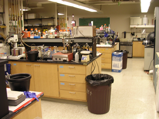
Although the main focus of our group is spectroscopy, many wet lab techniques are still used and are vital to our research.
We grow our own E. coli or yeast cultures that express proteins of interest,
then we purified the proteins and reconstitute them into E. coli polar lipids.
Site-directed mutagenesis is another major technique utilized in our lab.
Specific bases in the DNA are changed to study the effects of specific protein residue changes,
which helps us in determining roles of those specific residues.
Other techniques are also utilized, including pH titration, isotope labeling, and hydrogen-deuterium exchange.
Near-IR resonance Raman spectroscopy
Resonance Raman spectroscopy is an inelastic scattering vibrational spectroscopy that
takes advantage of signal enhancement when excitation is near a real absorption band.
We use a 785nm laser to excite our proteins (absorption ranging from 440nm to 570nm).
Although this is quite far from the actual absorption peak,
vibrational peaks from the source of the absorption (the chromophore, or the retinal molecule) are enhanced.
However, the laser is far enough from the absorption peak that it does not excite the protein.
This allows us to probe vibrational bands near the retinal in the protein's ground state
without the vibrational bands from the rest of the protein.
This technique gives us a clear picture of the ground state.
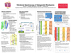
To see an example of resonance Raman spectroscopy results,
look at the GtACR1 poster recently presented at the 2016 Biophysical Society Meeting
by clicking on the image on the left.
Low-temperature FTIR difference spectroscopy
Fourier-transform infrared (FTIR) spectroscopy is a direct absorption method
that probes vibrational bands of the entire protein.
Although, this is not useful on its own, difference spectroscopy can be used to detect
changes beween two states of the protein as small as 1 in thousands of the absorbance intensity.
We utilize this to study the changes that occur between the ground state and an excited photointermediate state.
A liquid nitrogen cooled cryostat is used to cool samples to cryo-temperatures
to drive proteins to specific photointermediate states of interest.
We can detect changes in retinal conformational state, hydrogen bonding changes, protonation state changes, and more.
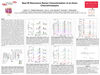
To see an example of low-temperature FTIR difference spectroscopy results,
look at the CaChR1 poster presented at the 2015 Biophysical Society Meeting
by clicking on the image on the left.
Time-resolved difference spectroscopy
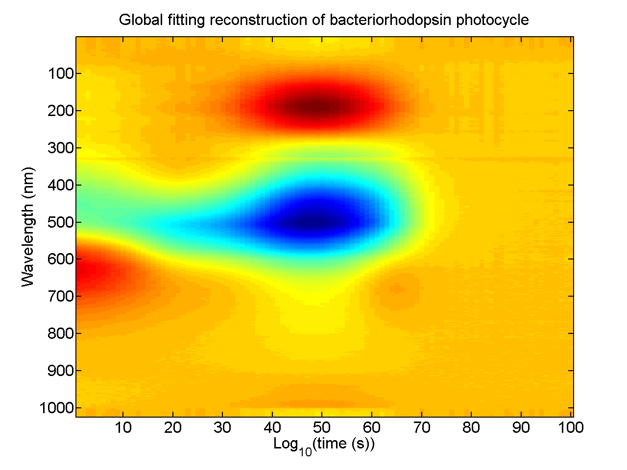
Our lab has capabilities to do time-resolved spectroscopy in both visible and infrared regions.
For both systems, a high-power nanosecond pulse laser is used to excited the sample,
which sits in a temperature controlled holder.
A pre-laser spectrum is subtracted from series of spectra acquired after the laser flash.
The visible spectrometer is a home-built system, with a fiber optic-connected Xenon flash lamp as the probe light source.
The timing between the laser and the Xenon flashes are controlled through a digital delay generator to map out the time axis.
The FTIR spectrometer uses a customized Bruker IFS 66v/S system.
Two styles of scans can be implemented with this system:
rapid scan, which requires shorter total acquisition time (several hours) but has lower time-resolution (~8ms),
and step scan, which requires longer total acquisition time (several days) but has higher time resolution (~1μs).